
The individual Excellence Fellowships – each of which is endowed with 350,000 euros – are going to the following eight candidates:
PD Dr. Paul Bröckelmann, Dept. of Internal Medicine I, University Hospital Cologne and Max Planck Institute for Biology of Ageing, Cologne
Project: Identification of immunotherapeutic vulnerabilities in tumor ecosystems of B cell lymphomas
Lymphomas are a heterogeneous group of hematological malignancies that merely partially and frequently only temporarily respond to currently available immunotherapies. The research project aims to characterize the immune landscape of patients with lymphomas in detail with the help of multifaceted modern analysis techniques and genetic screening methods. The overarching objective is the identification of mechanisms for the response or the failure to respond to immunotherapy in order to develop new treatment strategies in this manner.
PD Dr. Markus Eckstein, Institute for Pathology, Erlangen University Hospital, FAU Erlangen-Nuremberg
Project: Decoding immunotherapy resistance mechanisms in the case of locally advanced and metastasized urothelial carcinomas
Muscle-invasive and metastatic bladder cancer belong to the most aggressive types of cancer. Despite new therapies such as immunotherapy, in recent decades the survival rates for this type of cancer have not improved significantly. It is presently not possible to predict via biomarkers which patients benefit from modern immunotherapies. Moreover, to date we do not understand to a sufficient extent why this type of cancer is so resistant to immunotherapies. Within the planned project it is therefore intended to research more intensively how bladder cancer evades the immune system and evolves during the course of metastasis in order to develop novel, tailored immunotherapeutic approaches for patients with muscle-invasive and metastasized bladder cancer.
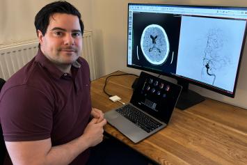
PD Dr. Tobias Faizy, Dept. of Interventional Neuroradiology, Münster University Hospital
Project: Development of a new, image-based diagnosis concept for assessing patients with an acute stroke – Improvement of patient selection and prediction of the clinical outcome
During an ischemic stroke the smallest blood vessels, so-called collateral vessels, can temporarily supply brain tissue in danger of dying. These collateral vessels bear an extreme importance in stroke therapy. Within the scope of this project a new concept for structural diagnosis and the quantification of the brain’s collateral vessels are supposed to be developed. The studies are based on datasets stemming from computed tomography that are generated by means of artificial intelligence (AI).
PD Dr. Julian Mustroph, Dept. for Internal Medicine II, University Hospital Regensburg
Project: Cellular pathophysiology of human cardial TTR amyloidosis
In the course of transthyretin (TTR) amyloidosis abnormally folded proteins, so-called transthyretin amyloid, are deposited in the heart. The disease can occur due to physical aging or a genetic defect; the resulting protein deposits lead to a thickening and stiffening of cardiac muscle. Within the scope of the research project small human heart samples from so-called myocardial (heart muscle) biopsies and the living myocardial cells taken from them are intended to be utilized for the first time and examined.
PD Dr. Alexander Nave, Dept. of Neurology and Experimental Neurology, Charité – University Hospital Berlin
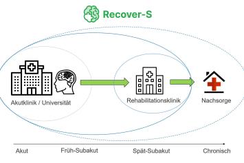
Project: Cross-sectoral studies following a stroke – Predictors of stroke recovery in personalized stroke rehabilitation
Recovery following a stroke varies greatly. The cross-sectoral index named RECOVER-S studies patients’ neurological and psychological recovery following a stroke and enables an extensive collaboration between the various actors involved in stroke care. Biomarkers indicating recovery following a stroke are supposed to be identified and validated via MRI scan examination and blood tests. The influence of wearable sensors on the motivation and recovery progress of stroke patients is intended to be studied for the first time within an index-based clinical trial.
PD Dr. Jan Peeken, Dept. of Radiation Oncology, University Hospital rechts der Isar, Technical University Munich (TUM)
Project: Individualized risk assessment using AI and molecular imaging for prostate cancer patients
The development of artificial intelligence (AI) provides new possibilities for the analysis of medical image data. For example, AI algorithms are able to analyze tumors on a non-invasive basis and generate prognoses. In association with a group of international medical treatment centers the researchers compile data on patients with prostate cancer who have received a specific form of molecular imaging (PSMA PET) prior to radiooncological treatment. The development of specialized AI algorithms is intended to improve therapy planning, characterize tumors better and support physicians during prognostic assessment. The models developed are ultimately supposed to be tested in the course of a clinical trial.
PD Dr. Adrian Regensburger, Project Team Translational Pediatrics, Pediatric Gastroenterology and Pediatric Ultrasound Imaging, Dept. of Child and Adolescent Medicine, University Hospital Erlangen
Project: Translational optoacoustic biomarkers for diseases of the gastrointestinal (GI) tract
While the treatment of children and adolescents is becoming more and more complex and structures are being addressed on an increasingly smaller level down to the genes themselves, physicians often lack suitable instruments toward assessing therapy success. Innovative, non-invasive imaging techniques such as optoacoustics might be able to provide corresponding molecular tissue information in the future, even in the case of the smallest patients. In addition, by means of this ultrasound imaging that displays molecular sensitivity findings from experiments on animals can be translated directly into clinical application. Within the scope of the Excellence Fellowship the researchers are going to develop novel diagnostic areas for optoacoustics in the case of intestinal and liver diseases in the course of translational trials. The aim is to establish optoacoustic imaging in clinical diagnostics for children and adolescents, and thus spare them invasive procedures.
PD Dr. Eva Schrezenmeier, Dept. of Nephrology and Medical Intensive Care, Charité – University Hospital Berlin
Project: More effective therapies for immune-mediated renal diseases through a deeper understanding of the development of B cells and plasma cells
Immune-mediated renal (i.e. kidney) diseases vary greatly in the ways they progress, from mild and self-limiting all the way to severe and insufficiently treatable. B and plasma cells play a key role in their pathogenesis. The aim of this project is to research the differences in the formation and maintenance of B-cell and plasma-cell memory between various courses of progression in immune-mediated renal diseases and derive new therapies from them.