Imaging of immune responses and tumor cell invasion in glioma models
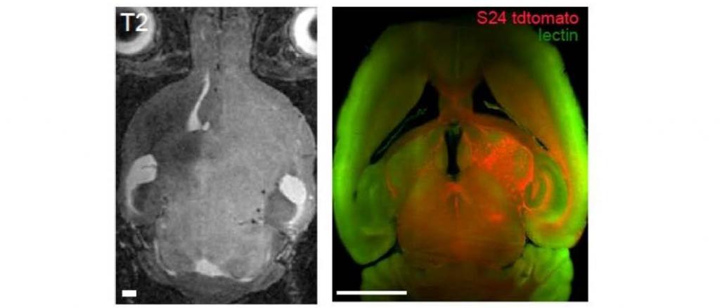
Glioblastoma is a malignant brain tumor with poor survival. Imaging in glioblastoma, mainly performed by magnetic resonance imaging, is restricted to morphological information (tumor size, perifocal edema). Immunotherapies aim to elicit anti-tumor immune responses but monitoring treatment effects beyond the macroscopic tumor size remains difficult. Visualizing immune responses by direct tracking of effector cells could facilitate preclinical therapy development and allow treatment monitoring and patient stratification. We aim to develop imaging tools to combine MRI and optical methods to visualize tumor cell infiltration as well as innate and adaptive immune responses that are mounted by immunotherapeutic treatment regimes.
Here you can get further information.
