
Particularly at the start of their medical career, physicians face the challenge of combining clinical responsibilities with scientific research. This year, nine early-career researchers will receive a two-year Else Kröner Memorial Fellowship, each endowed with €250,000. A fellowship allows recipients to take time away from their clinical responsibilities to focus fully on advancing promising medical research. At the same time, it lays the foundations for a career as a clinician scientist.
The following physicians are to be awarded a fellowship:
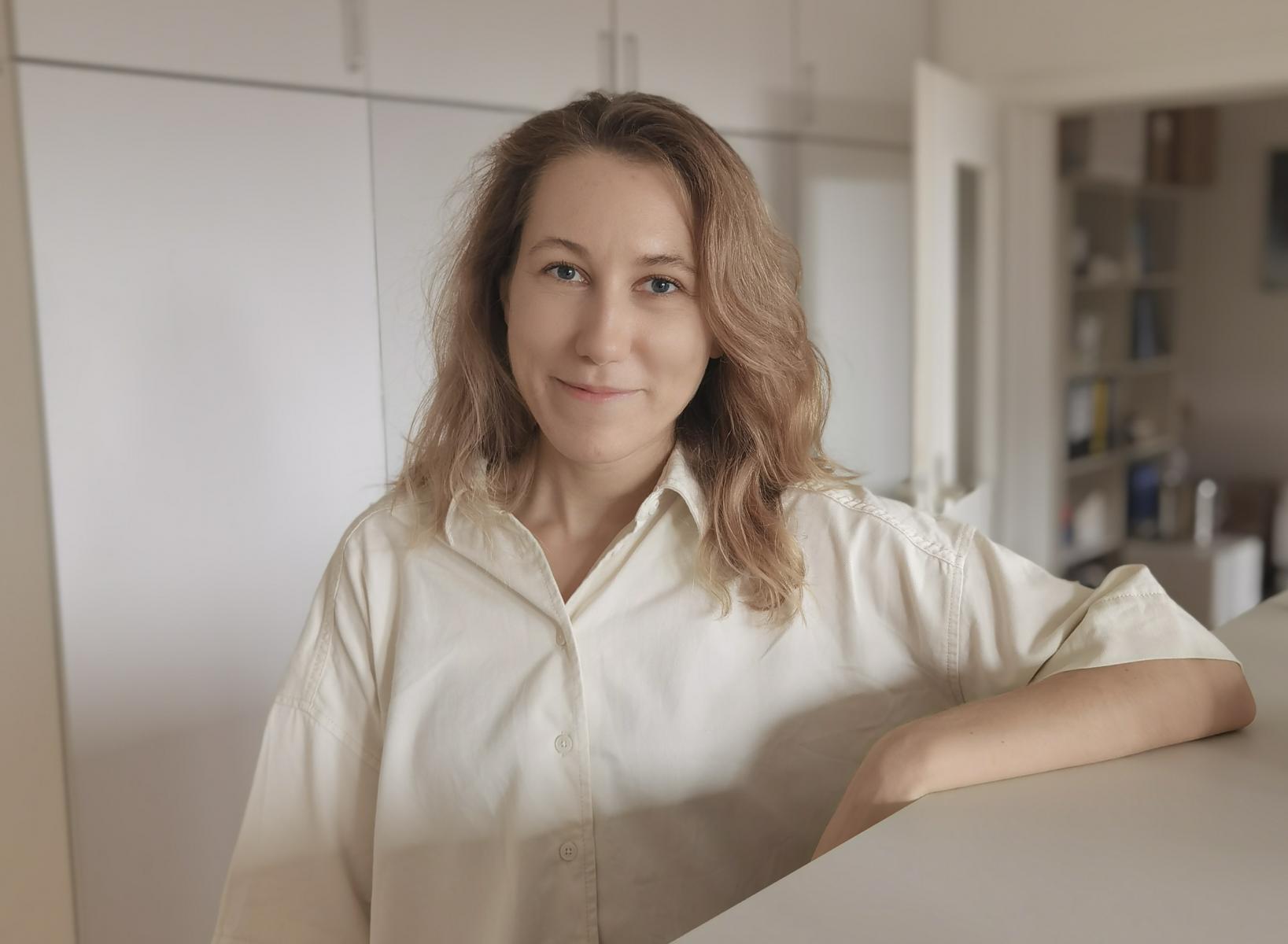
Dr. Ekaterina Friebel, Institute of Neuropathology, Charité – Universitätsmedizin Berlin
Project: Cellular and Transcriptomic Changes Driving Differences in the Clinical Phenotype of Amyotrophic Lateral Sclerosis (ALS)
Amyotrophic lateral sclerosis (ALS) is an incurable disease in which the nerve cells that control muscle movement gradually die. The course of the disease can vary, however: In some patients, nerve cells in the brain are primarily affected, while in others, degeneration is more pronounced in the brainstem and spinal cord. In this project, Dr. Ekaterina Friebel examines tissue from different regions of the brain and spinal cord of the same ALS patients. She uses state-of-the-art single-cell techniques to investigate changes in nerve cells and their supporting cells, with a particular focus on microglia. The aim is to understand why ALS progresses differently between patients and to identify markers that could enable more precise diagnosis.
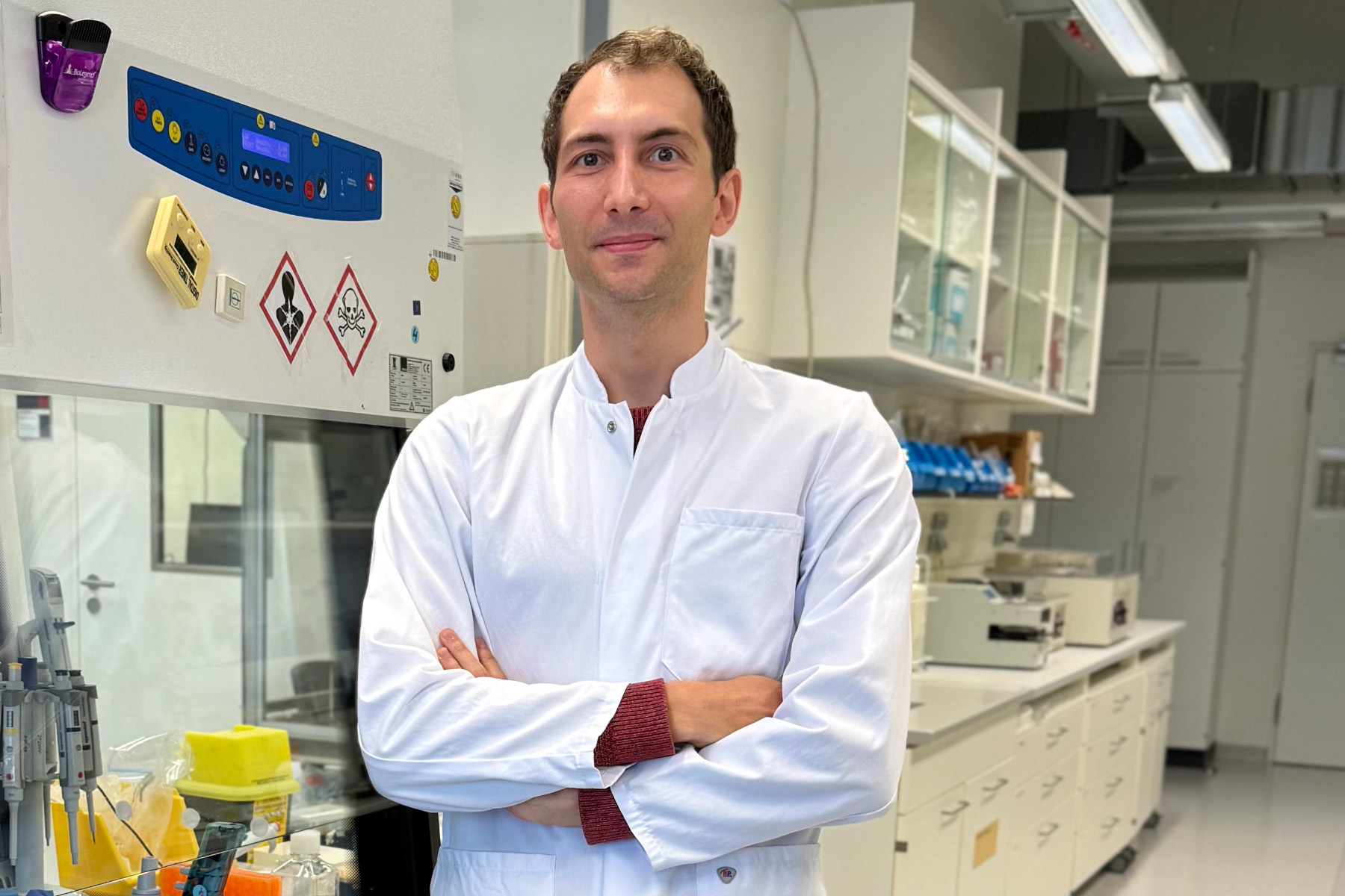
Dr. Maximilian Haist, Department of Dermatology, University Medical Center Mainz
Project: Perturbing the Spatial Immune Landscape in Merkel Cell Carcinoma to Prevent Distant Metastasis Formation
Merkel cell carcinoma (MCC) is a rare but aggressive skin cancer with a high rate of metastasis and a poor prognosis. Disease progression is influenced by infection with Merkel cell polyomavirus (MCPyV) and by UV-induced DNA damage. Dr. Maximilian Haist and his team are developing an atlas of the MCC tumor microenvironment to map tumor-immune interactions and identify features of virus-associated immunomodulation associated with the risk of distant metastatic progression. The findings will be validated in independent external MCC cohorts to improve patient stratification and treatment selection.
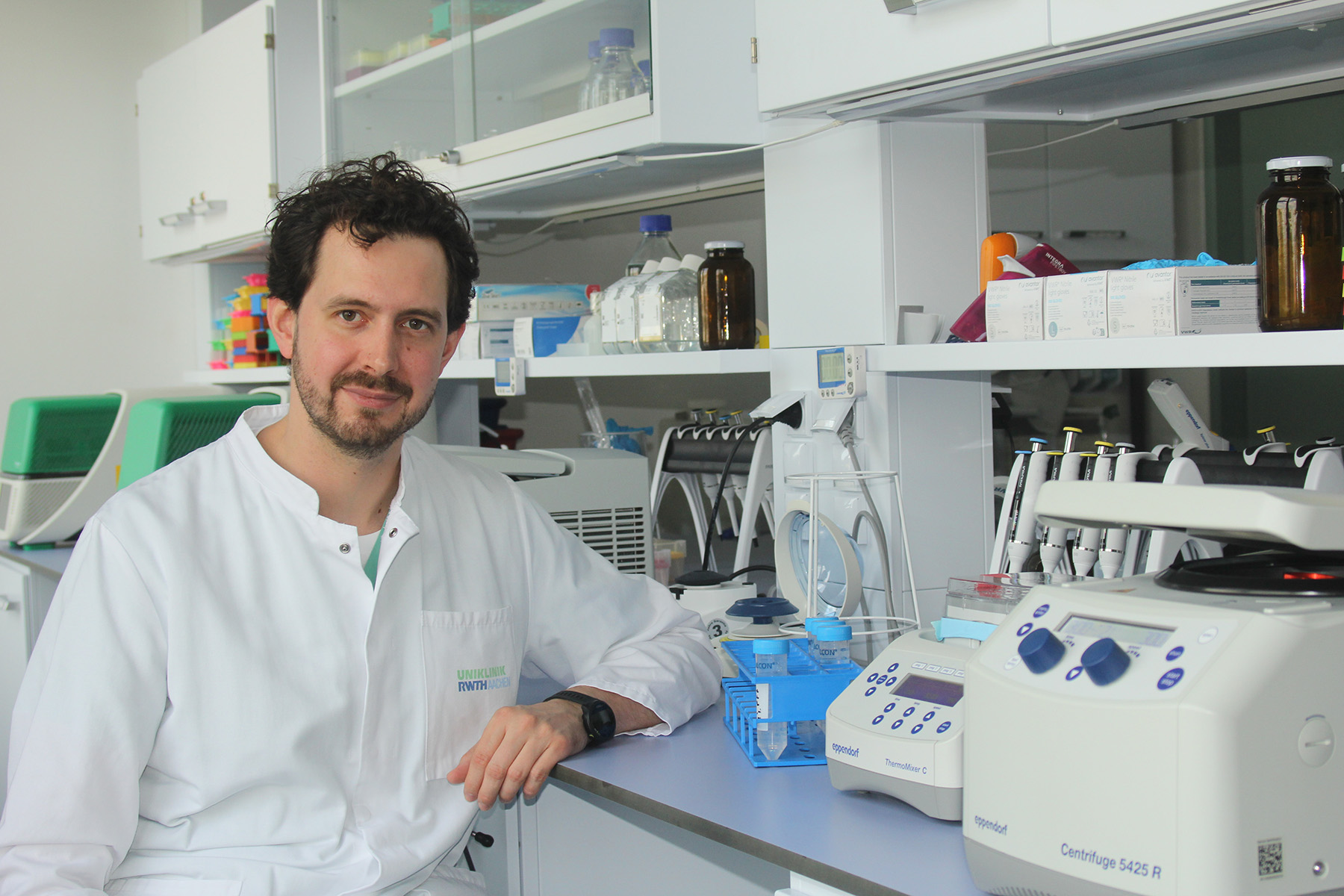
Dr. Konrad Hoeft, Department of Medicine 2 (Nephrology, Rheumatology, Clinical Immunology and Hypertension), RWTH Aachen University
Project: Interrogating Chronic Kidney Disease and Fibrosis as Drivers of Heart Failure with Preserved Ejection Fraction
Many people with chronic kidney disease develop a particular type of heart failure in which the heart continues to pump normally, but becomes stiff and can no longer fill properly with blood. It is not yet fully understood why diseased kidneys alter the heart. One possible explanation is that waste products accumulate in the blood and cause fibrosis of the heart tissue. Dr. Konrad Hoeft is examining diseased heart tissue at the cellular level and testing his findings using miniature hearts grown from stem cells. The goal is to better understand how the kidneys and heart interact and to identify new treatment approaches.
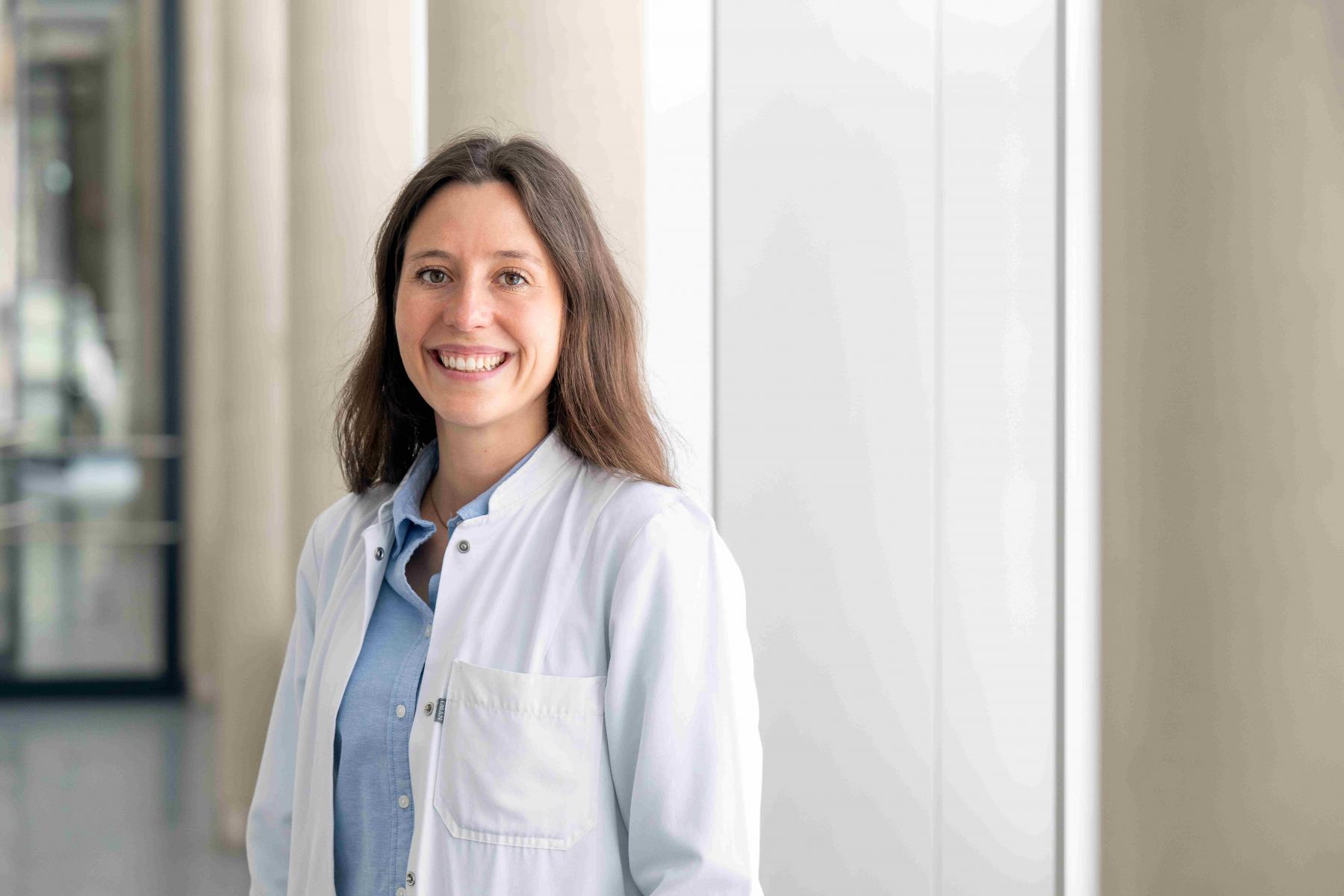
Dr. Friederike Jungmann, Institute for interventional and diagnostic Radiology, TUM University Hospital
Project: From Benchmarks to Bedside: Improving Safety and Reliability of LLM-Based Clinical Decision Support with Evidence-based Medical Reasoning
AI is increasingly being integrated into many areas of our lives, including medicine. This project brings together different approaches for integrating medical “knowledge” into the latest large language models (LLMs) and assessing their clinical impact. It aims to strengthen the LLMs’ clinical reasoning capabilities; enable robust, data-driven evaluation; and support the development of a medically grounded clinical decision-support system. By improving the robustness, medical accuracy, and adherence to clinical guidelines of LLMs, the project aims to enhance patient safety in an increasingly digital healthcare system.
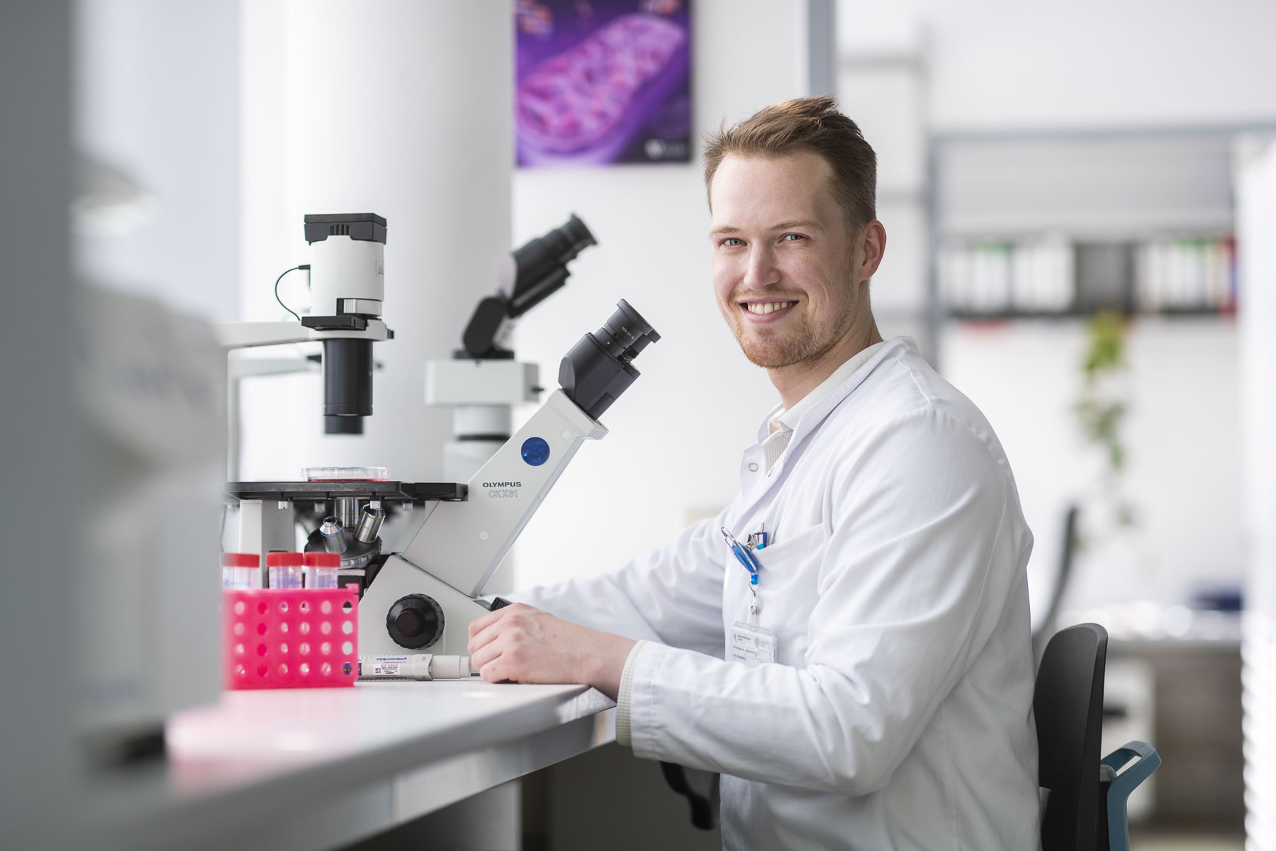
Dr. Benjamin Klein, Department of Dermatology, Venerology and Allergology, Faculty of Medicine, University of Leipzig
Project: From Molecular Signatures to Personalized Therapy in Cutaneous Lupus Erythematosus
Cutaneous lupus erythematosus (CLE) is a chronic inflammatory skin disease characterized by substantial clinical and molecular heterogeneity. Established therapies are not equally effective for all patients. This project will investigate how molecular signatures can help characterize disease activity and treatment response. To this end, clinical data will be combined with molecular analyses of skin samples, functional studies using primary cells, and longitudinal analyses of immune cells in the blood to track changes in the disease course and response to treatment. The goal is to identify molecular endotypes that enable a more accurate assessment of individual disease risk and more personalized treatment decisions for patients with CLE.
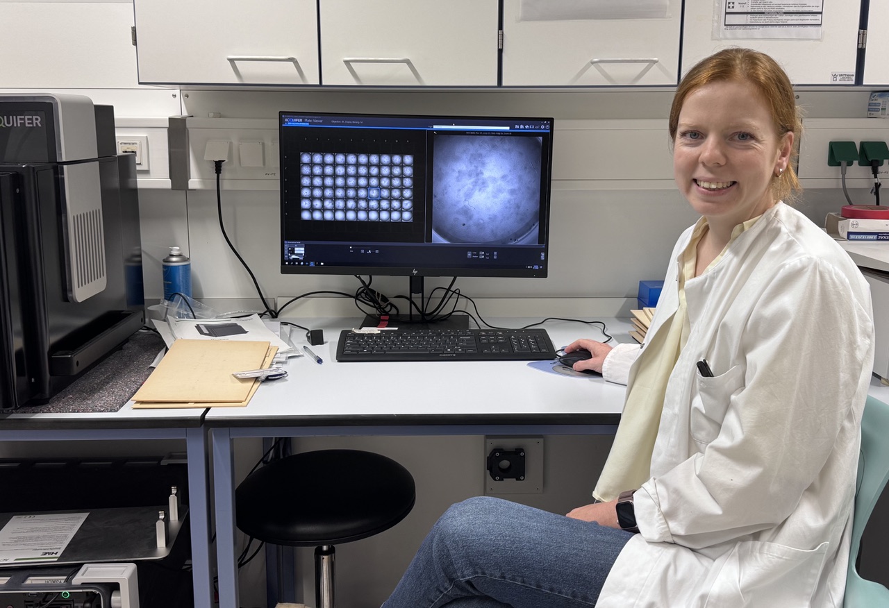
Caroline M. Kolvenbach, Center for Pediatrics and Adolescent Medicine, University Hospital Heidelberg
Project: Deciphering Allele-Dependent Phenotypic Divergence in HNF1B-Associated Kidney Disease
Genetic changes in the HNF1B gene are a common cause of congenital kidney malformations. The types of malformations and extent of kidney function impairment can vary considerably, however. In this project, Caroline M. Kolvenbach and her team investigate how different variants of this gene affect kidney development and function. The findings will help to better understand the diverse manifestations of the disease and identify possible future treatment approaches.
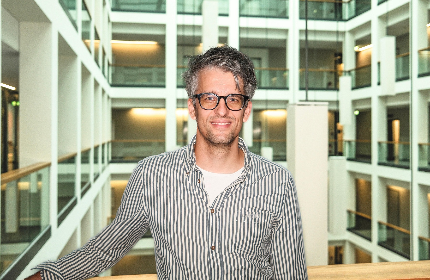
Dr. Jakob Kreye, Department of Pediatric Neurology and Institute of Cell Biology and Neurobiology, Charité – Universitätsmedizin Berlin
Project: Maternal Autoantibody Signatures in Autism Spectrum Disorders
Autism spectrum disorders affect around one percent of children worldwide. Beside genetic risk factors, maternal autoantibodies may also play a role. These antibodies can cross the placenta to the unborn child and affect its developing nervous system. Dr. Jakob Kreye and his team examine blood samples from the mothers of children with autism and control participants to identify relevant autoantibodies and the structures they target. The aim is to determine their frequency and characteristics. In the long term, these findings could lead to a better understanding of the biological mechanisms underlying autism and provide a basis for new biomarkers and targeted medical approaches.
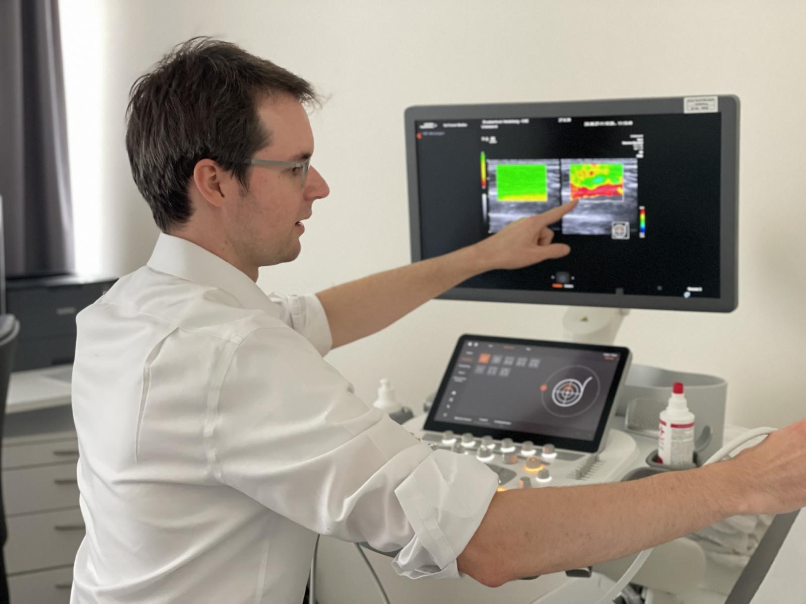
Dr. André Pfob, Institute for Medical Artificial Intelligence (IMAI), Heidelberg University Hospital and Faculty of Medicine, University of Heidelberg and Breast Center Heidelberg at St. Elisabeth Clinic
Project: Artificial Intelligence Assisted Shear-Wave Elastography for Improved Breast Cancer Diagnosis: A Prospective Phase II Validation Study
Ultrasound is an important diagnostic tool for evaluating suspicious breast lesions. However, because benign and malignant lesions can look similar, many women undergo biopsies that ultimately reveal no cancer. While shear-wave elastography provides additional information by measuring tissue stiffness, the results can be difficult to interpret. Dr. André Pfob and his team have therefore developed an AI-based method that automatically analyzes elastography images. In preliminary studies, the model reliably detected breast cancer, reducing unnecessary biopsies. The Else Kröner Memorial Fellowship will enable evaluation of this approach for its prospective use in routine clinical practice for the first time.
Dr. Sebastian Ziegelmayer, Institute for Interventional and Diagnostic Radiology, TUM University Hospital
Project: Imaging-based Modeling of the Tumor-Macroenvironment in Gastrointestinal Cancer
Malignant tumors do not act in local isolation, but rather can alter the entire body – the metabolism, muscles, fatty tissue, and internal organs, for instance. These systemic changes in turn influence the course of disease, treatment tolerance, and the development of metastases. This project uses routine CT scans from patients with gastrointestinal tumors to investigate the tumor macroenvironment. An AI model automatically maps organs and tissues and analyzes how they change over time. The goal is to predict overall survival, patterns of metastasis, and treatment toxicity, thereby enabling more precise disease stratification.